D2IMDeformation from greyscale
Predicts bone deformation directly from X-ray tomography greyscale, without an explicit correlation step.
Extreme Mechanics Letters10.1016/j.eml.2024.102202Predictive Imaging Lab, School of Engineering, University of Greenwich
Decoding biological tissues and bio‑inspired materials with in‑situ X-ray microscopy, full-field measurement, artificial intelligence and quantum insight.
Bone, soft tissue, bio-composites and self-assembled biomaterials: imaged under load, then modelled.
A tomogram is a measurement, not a picture. We build the models that read it.
The Predictive Imaging Lab is led by Gianluca Tozzi, Professor of Industrial Engineering at the University of Greenwich. The group advances the understanding of biological tissues and biomaterials by integrating advanced imaging (in‑situ X-ray tomography, hyperspectral imaging) with full-field measurement such as digital volume correlation, and with artificial intelligence.
Named models from the lab, with the papers behind them.
Predicts bone deformation directly from X-ray tomography greyscale, without an explicit correlation step.
Extreme Mechanics Letters10.1016/j.eml.2024.102202Improves classification accuracy in hyperspectral imaging using a diffusion-based representation.
Scientific Reports10.1038/s41598-024-58125-4Improves tumour detection from hyperspectral images of biological tissue.
Journal of Microscopy10.1111/jmi.13372Six programmes, each led by a member or affiliate of the lab.
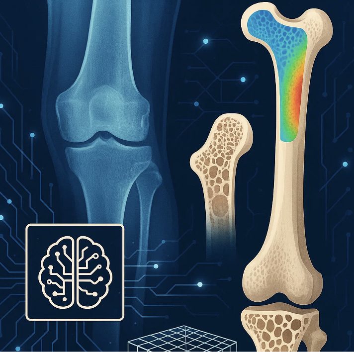
Fully developing D2IM by integrating additional AI strategies, advanced XCT segmentation models and the power of large language models.
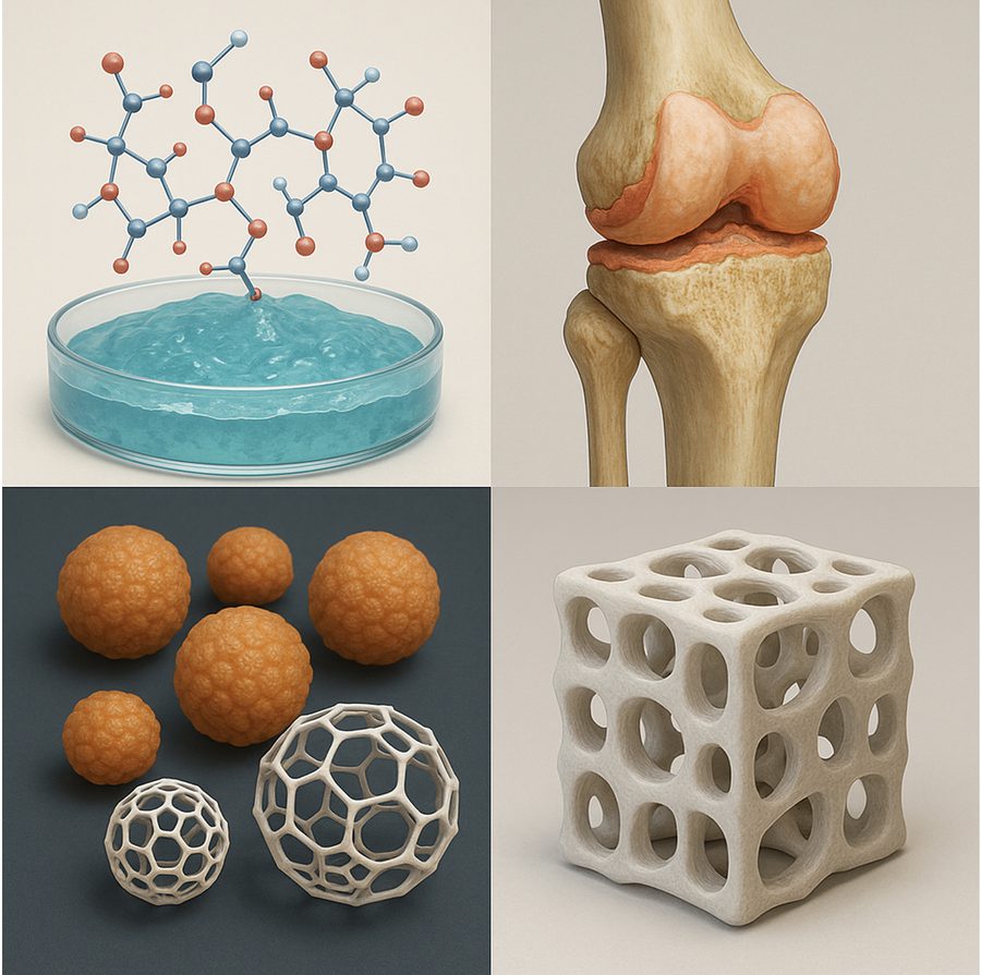
Applying advanced AI models to analyse, predict and optimise the hierarchical self-assembly of nanoparticles into macroscale soft biomaterials.
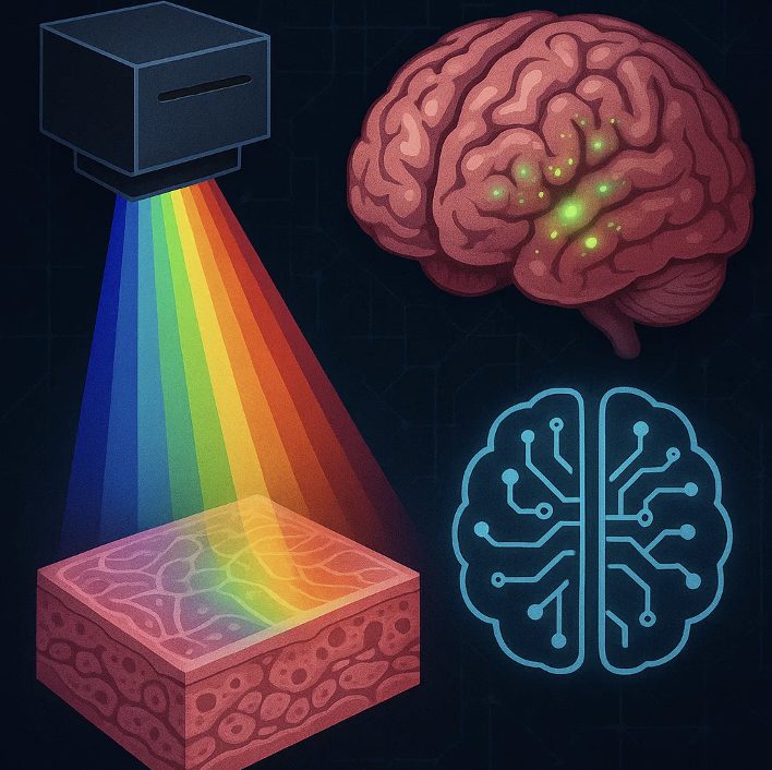
Exploring the unique spectral signature of biological tissues and developing advanced AI models for healthcare.
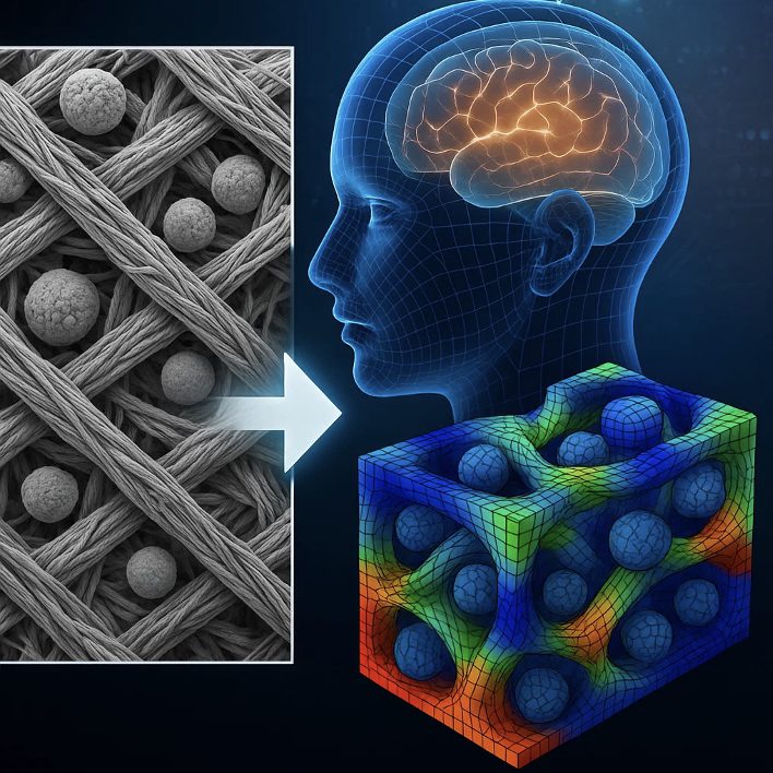
Developing D2IM-based tools that give the industrial partner a technology to identify the nature of defects in bio-composites and inform manufacturing.
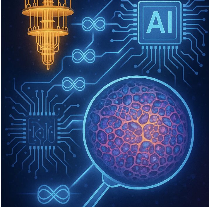
Developing quantum-native representations of biological tissue images, enabling efficient classification, segmentation, measurement and multimodal fusion.
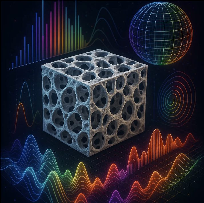
A cross-faculty collaboration investigating AI sonification to design and optimise bone biomimetic materials, blending imaging, computational modelling and additive manufacturing.
Faculty of Engineering and Science, University of Greenwich.
KA Imaging
Sub-micron resolution with fast phase-contrast acquisition, paired with a custom-designed in‑situ loading device so specimens can be imaged while they deform.
Living Optics
Spectral signatures that reveal the biochemical state of tissue alongside its structure, and feed our HSI classification and tumour-detection models.
We take on PhD researchers, postdocs and industrial partners working at the intersection of imaging, mechanics and machine learning. Bring a specimen, a dataset or a question.